
CORONECTOMY: WHAT ARE THE STEPS FOR THE CLINICIAN TO PERFORM THIS TECHNIQUE? – DESCRIPTION OF THE SURGICAL TECHNIQUE
Augusto Dutra Vasconcellos1, Peter Reher2, Júlia Eick Iglesias3, Gustavo Vicentis Oliveira Fernandes4,5*, Marcelo José Uzeda1,6, Rodrigo Figueiredo de Brito Resende1,6
1 School of Dentistry, Universidade Iguaçu, Nova Iguaçu, RJ, Brazil.
2 School of Dentistry, The University of Queensland, Brisbane, QLD, Australia.
3 School of Dentistry and Medical Sciences, Charles Sturt University, Orange, NSW, Australia.
4 School of Dentistry and Oral Health, A.T. Still University, St. Louis, MO, USA.
5 Interdisciplinary Centre for Investigations in Health, Universidade Católica Portuguesa, Viseu, Portugal
6 School of Dentistry, Universidade Federal Fluminense, Niterói, RJ, Brazil.
*
Corresponding author: Gustavo Vicentis Oliveira Fernandes E-mail : gustfernandes@gmail.com
ABSTRACT
Coronectomy, also described as intentional partial odontectomy, is an alternative surgical technique for the extraction of teeth that present an increased risk of injury to the Inferior Alveolar Nerve (IAN) and/or mandibular fractures. It is essential that the clinician correctly indicates the procedure and observes key intraoperative precautions to ensure success. The objective of this paper is to provide a step-by-step description of this surgical technique and to outline its proper indications and contraindications, enabling clinicians to achieve a high success rate with this procedure. A search was performed in the PubMed/MEDLINE databases using the keywords: “Odontectomy,” “Third Molar,” and “Coronectomy.” The inclusion criteria were English-language articles published between 1984 and 2023 that addressed coronectomy as an alternative to mandibular third molar extraction. In addition, literature review articles evaluating the feasibility and effectiveness of the technique were included. Exclusion criteria included studies that did not address coronectomy or discuss its efficacy and safety, as well as those that focused solely on the extraction of third molars without the use of coronectomy.
Keywords: Odontectomy, Third Molar, Coronectomy.
INTRODUCTION
Extraction of third molars and impacted supernumerary teeth is one of the most requested procedures in daily dental practice, and it is frequently accompanied by common signs and symptoms such as pain, bleeding, trismus, or even infections (pericoronitis). To perform this procedure, accurate planning is necessary to minimize the risks of injury to the inferior alveolar nerve (IAN), root fractures, or even mandibular fractures.1-3 Coronectomy, also termed intentional partial odontectomy in the literature, consists of sectioning the coronal portion of the tooth while allowing the roots to remain intact within the dental socket in situ, in order to minimize damage to the IAN and/or mandibular fractures. This technique was first described by Ecuyer and Debien in 1984 for molars in proximity to the mandibular canal, providing greater patient safety. Although numerous studies have highlighted the effectiveness of the technique, it still presents negative aspects, such as susceptibility to infections due to the retained roots.4-7 The object of this paper is to outline the main aspects of coronectomy by describing its indications, precautions, and contraindications, as well as evaluating its effectiveness in avoiding temporary or permanent IAN complications, infections, or bone- related events.
METHODS
A search was carried out in the PubMed/MEDLINE databases using the keywords “Odontectomy,” “Third Molar,” and “Coronectomy.” The inclusion criteria were English- language articles published from 1984 to 2023 that addressed coronectomy as an alternative to the extraction of mandibular third molars. In addition, literature review articles that evaluated the feasibility and effectiveness of the technique were included. The exclusion criteria included articles that did not address coronectomy, that did not discuss its efficacy and safety, or that addressed only the extraction of third molars without the use of intentional partial odontectomy (Figure 1).
RESULTS
The database screening summary included PubMed/MEDLINE, yielding 69 articles. Fifty-two potentially eligible studies were found; 42 articles were excluded after analysis, and 22 articles were included.
Indications and contraindications of the technique:
Among the main criteria that must be considered for performing this technique are: the tooth must have pulp vitality, be in intimate proximity to the mandibular canal, and be impacted. Likewise, it must not present any carious lesions, periapical lesions, prior infections, mobility, or pain. The procedure is contraindicated when cysts or tumors are present. Some authors state that the organism can recognize root remnants of teeth with infections or mobility as foreign bodies.8-12 According to several studies, the technique is not indicated for teeth with caries at risk of pulp involvement, mobility, apical lesions, or the presence of cystic tissue. Moreover, it is not recommended for patients scheduled for esthetic-functional mandibular osteotomies, immunocompromised patients, or those who have previously received radiotherapy. Conversely, it is indicated for teeth with pericoronitis, those causing periodontal disease in adjacent teeth, or those that pose a risk of mandibular fracture.1-3,10,13,14 Intentional partial odontectomy may also cause postoperative infections, especially when the dental follicle is not completely removed upon crown sectioning. Approximately 10–12% of cases may be prone to infection.1,15-17
Advantages and disadvantages:
When evaluating the patient, it is necessary to identify the main risk factors for nerve injury, such as advanced age, mesiodistal angulation of the tooth (distoangular), lingual angulation of the tooth, lack of integrity of the lingual cortical plate, distal bone removal adjacent to the third molar, and long-standing pericoronitis that has resulted in soft tissue fibrosis. Third molar removal surgeries account for the majority of nerve injuries associated with extractions, most frequently involving the Inferior Alveolar Nerve (IAN) and the Lingual Nerve (LN). Among the most commonly cited complications are temporary paresthesia and permanent paresthesia.8,18,19
Neurological sequelae should be assessed as hypoesthesia, hyperesthesia, and dysesthesia of the lower lip and the mental region on the operated side when removing sutures. In addition, alterations in lingual sensitivity should be evaluated (Renton et al.). Although coronectomy does not completely eliminate the possibility of neurological damage and, in some instances, may require a secondary surgical stage for the removal of retained roots, it offers greater safety with respect to the IAN.18
Description of the surgical technique:
Imaging assessment:
A thorough imaging exam should be performed by means of cone-beam computed tomography (CBCT) to accurately assess the relationship between the dental roots and the mandibular canal and to support proper surgical planning.
Choice of local anesthetic:
For local anesthesia, 4% Articaine with 1:100,000 epinephrine or 2% Lidocaine with 1:100,000 epinephrine is preferred to achieve an appropriate regional nerve block and improved hemostasis.
Incision and flap elevation:
Make an incision with a no.15 (or 15c) blade mounted on a no. 3 or no. 7 handle to design a preferably triangular flap. Begin the incision from the most posterior tooth, along the alveolar crest distally, extending to the mesiobuccal line angle, with a vertical releasing incision to the alveolar mucosa. Elevate a full-thickness flap (mucosa, submucosa, and periosteum) using a periosteal elevator, such as a Molt no. 9 elevator (Figure 2).
Osteotomy:
Perform ostectomy with a surgical carbide round bur (no. 6 or no. 8) to expose the coronal portion of the tooth down to the cementoenamel junction, using a rotary instrument under constant and abundant irrigation with sterile 0.9% NaCl solution (Figure 3).
Odontosection:
Subsequently, using a Zekrya bur or a long shank 702 bur, perform the odontosection/odontectomy with buccolingual strokes, always respecting two-thirds of the buccolingual width of the tooth to avoid damage to the lingual nerve (Figure 4). For easier removal, a crown cut along the long axis of the tooth may be performed (Figure 5).
Crown removal:
Finally, using a Straight Seldin no. 2 elevator or an apex elevator no. 303, complete the coronectomy. If necessary, the elevator may be supported on the alveolar crest to gain a fixed fulcrum, avoiding contact with the roots (Figure 6). It is important to remember that the clinician should not mobilize the root portion, but only the coronal portion.
Bone adjustment:
Next, using a surgical carbide round bur no. 6, regularize the remaining roots by removing sharp edges, ensuring that they remain 3–4 mm below the alveolar crest (Figure 7).
Wound closure:
For wound closure, first place a simple interrupted suture at the flap apex and then along the entire incision line, thus avoiding postoperative exposure of the surgical site and/or dehiscence of the flap (Figure 8).
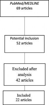
Figure 1. Flowchart of the literature review.
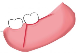
Figure 2. Creation of a triangular surgical flap to provide complete visualization of the region.
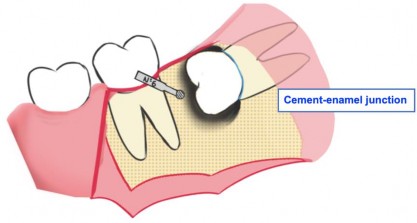
Figure 3. Ostectomy using a surgical carbide round bur to properly expose the crown of the impacted tooth.
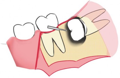
Figure 4. Odontosection with a surgical Zekrya bur for a buccolingual section, respecting two-thirds of this distance.
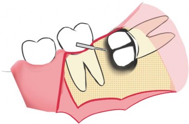
Figure 5. Odontosection with a surgical Zekrya bur for a mesiodistal section, respecting two thirds of this distance, facilitating complete crown removal with minimal risk of root movement.
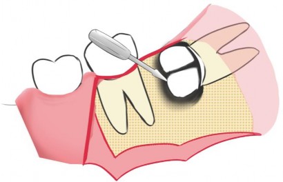
Figure 6. Separation and complete removal of coronal fragments using a Straight Seldin number 2 elevator.
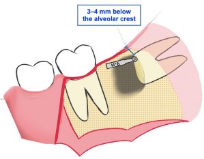
Figure 7. Ostectomy with a surgical round bur number 6 to regularize root remnants by
removing sharp edges, ensuring 3–4 mm below the bony margin.
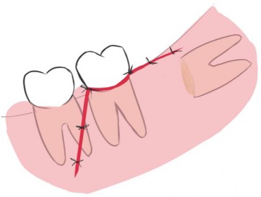
Figure 8. Closure of the triangular flap with 3-0 or 4-0 suture.
DISCUSSION
Third molar surgeries have been the aim of many studies, looking for a better post-operative outcome20,21 or providing advice about instruments and care.22,23 Corenectomy was first
described by Ecuyer and Debien in 1984 as a technique in which the roots are intentionally left within the dental socket after coronal sectioning.10 According to Williams et al.,24 panoramic radiography is widely used to assess the degree of proximity between an impacted tooth and the mandibular canal for the extraction of mandibular third molars; however, it may be more challenging to interpret. Therefore, cone-beam computed tomography (CBCT) offers greater visualization and safety for the procedure. Currently, it is indispensable to use CBCT in cases where the panoramic radiograph suggests intimate contact between the roots and the mandibular canal, as it provides greater precision without distortion in a three-dimensional view of the region.
Although coronectomy offers greater safety by avoiding potential IAN injury, the technique itself does not completely eliminate the possibility of neurological damage.10,18,24 Roots tend to migrate toward the superior border of the mandible during healing, and a further procedure to remove the retained roots may be necessary. There is also concern about the possibility of retained roots developing a radicular cyst, indicating a need for preventive removal. In addition, as noted by Monaco et al.,10 there is concern regarding pulp complications; therefore, the technique is not indicated for third molars affected by caries, pulp infections, or mobility. Monaco et al.7 reported that access should be achieved via a full-thickness mucoperiosteal flap, preferably with an anterior vertical incision placed distal to the mandibular second molar and extending along the external oblique ridge. The flap design should be related to the type of impaction. However, a triangular flap with a releasing incision distal to the second molar is more effective for primary-intention healing than an envelope flap. In a case series of 43 coronectomies for impacted third molars, root migration was most likely to occur within the first 3 months and decreased markedly after 6 and 12 months. Patients who exhibited migration within 3 months were younger than those who did not. Subsequently, in 2015,10 the same authors determined that the retained roots should remain at least 3 mm below the lingual and buccal bony margins. Kim HS et al.25 and Renton et al.8 followed the same protocol, setting the cut margin 3 mm below the adjacent bone after crown removal. Mobility of the remaining roots was assessed to minimize secondary trauma to the roots.
For the technique, atraumatic removal of the coronal portion is performed by sectioning in the mesiodistal and buccolingual directions. The alveolar crest can be used as a support wall for elevators, thereby avoiding contact with the roots. Ostectomy may be necessary in some situations; however, it is recommended that it not be extended below the cementoenamel junction. By observing these details, stress on the roots is significantly reduced, as is the risk of technique failure and root mobilization during the procedure.26 According to Monaco et al.,10 the amount of bone to be removed must be compatible with the depth of inclusion and type of impaction observed. For vertical impactions or distoangular inclusions where 50% of the third molar crown is above the cementoenamel junction (CEJ) of the second molar, odontosection can be performed without ostectomy. However, if the crown is 50% or more below the CEJ of the second molar, ostectomy becomes necessary. For mesial or horizontal inclusions, ostectomy is more often required and should be performed until complete crown visualization is achieved. Regarding suturing, most studies indicate that suturing stabilizes the clot and improves postoperative healing. Suturing should provide tension-free healing, and interrupted simple stitches should be placed until the incision is completely closed, as depicted in Figure 7. Then, coronectomy is a valid surgical option for managing mandibular third molars. With adequate knowledge and attention to technique, the chances of success are high, and the incidence of potential complications is reduced. Proper surgical planning and mastery of the method are essential for the steps to be performed correctly and for success to be achieved.
REFERENCES
-
Mukherjee S, Vikraman B, Sankar D, Veerabahu MS. Evaluation of Outcome Following
Coronectomy for the Management of Mandibular Third Molars in Close Proximity to Inferior Alveolar Nerve. J Clin Diagn Res. 2016;10(8): ZC57–ZC62. doi: 10.7860/JCDR/2016/20991.8273
-
Cevera-Espert JC, Pérez-Martínez S, Cervera-Ballester J, Peñarrocha-Oltra D, Peñarrocha- Diago M. Coronectomy of impacted mandibular third molars: A meta-analysis and systematic review of the literature. Med Oral Patol Oral Cir Bucal. 2016;21(4):e505–e513. doi: 10.4317/medoral.21074
-
Barcellos BM, Velasques BD, Moura LB, Xavier CB. What Are the Parameters for Reoperation in Mandibular Third Molars Submitted to Coronectomy? A Systematic Review. J Oral Maxillofac Surg. 2019;77(6):1108–1115. doi: 10.1016/j.joms.2019.01.013
-
Moreno-Vicente J, Schiavone-Mussano R, Clemente-Salas E, Marí-Roig A, Jané-Salas E, López-López J. Coronectomy versus surgical removal of the lower third molars with a high risk of injury to the inferior alveolar nerve. A bibliographical review. Med Oral Patol Oral Cir Bucal. 2015;20(4): e508–e517.
-
Leung YY. Guided bone regeneration to reduce root migration after coronectomy of lower third molar: a randomized clinical trial. Clin Oral Investig. 2019;23(4):1595–1604.
-
Goto S, Kurita K, Kuroiwa Y, Hatano Y, Kohara K, Izumi M, Ariji E. Clinical and Dental Computed Tomographic Evaluation 1 Year After Coronectomy. J Oral Maxillofac Surg. 2012;70(5):1023–1029. doi: 10.1016/j.joms.2011.09.037
-
Monaco G, Santis G, Gatto MRA, Corinaldesi G, Marchetti C. Coronectomy: A Surgical Option for Impacted Third Molars in Close Proximity to the Inferior Alveolar Nerve. J Am Dent Assoc. 2012;143(4):363-369. doi: 10.14219/jada.archive.2012.0178
-
Renton T, Hankins M, Sproate C, McGurk M. A randomized controlled clinical trial to compare the incidence of injury to the inferior alveolar nerve as a result of coronectomy and removal of mandibular third molars. Br J Oral Maxillofac Surg. 2005;43(1):7–12. doi: 10.1016/j.bjoms.2004.09.002
-
Long H, Zhou Y, Liao L, Pyakurel U, Wang Y, Lai W. Coronectomy vs. Total Removal for Third Molar Extraction: A Systematic Review. J Dent Res. 2012;91(7):659–665.
-
Monaco G, Vignudelli E, Diazzi M, Marchetti C, Corinaldesi G. Coronectomy of mandibular third molars: A clinical protocol to avoid inferior alveolar nerve injury. J Cranio- Maxillofac Surg. 2015;43(8):1694–1699. doi: 10.1016/j.jcms.2015.07.006
-
Samani M, Henien M, Sproat C. Coronectomy of mandibular teeth other than third molars: a case series. Br J Oral Maxillofac Surg. 2016; 54:791–795.
-
Nishimoto RN, Moshman AT, Dodson TB, Beirne OR. Why Is Mandibular Third Molar Coronectomy Successful Without Concurrent Root Canal Treatment? J Oral Maxillofac Surg. 2020;78(11):1886–1891.
-
Richards JA, Coyle MJ, Beech AN. Plain radiographs for the assessment of coronectomy: not always a case of black or white. Br J Oral Maxillofac Surg. 2019;57(7):663–665.
-
Matzen LH, Villefrance JS, Norholt SE, Bak J, Wenzel A. Cone Beam CT and treatment decision of mandibular third molars: removal vs. coronectomy — a 3-year audit. Dentomaxillofac Radiol. 2020;49(3):20190250. doi: 10.1259/dmfr.20190250
-
Sarwar H, Mahmood-Rao S. Coronectomy; Good or Bad? Dent Update. 2015;42(9):824– 828. doi:10.12968/denu.2015.42.9.824
-
Kang F, Xue Z, Zhou X, Zhang X, Hou G, Feng Y. Coronectomy: A Useful Approach in Minimizing Nerve Injury Compared with Traditional Extraction of Deeply Impacted Mandibular Third Molars. J Oral Maxillofac Surg. 2019;77:2221.e1–2221.e14. doi: 10.1016/j.joms.2019.06.186
-
Tuk J-G, Yohannes L-E, Ho J-T, Lindeboom J-A. Oral health-related quality of life after coronectomy for impacted mandibular third molar in the first postoperative week.
Med Oral Patol Oral Cir Bucal. 2021;26(5):e561–e567. doi: 10.4317/medoral.24506
-
Uzeda MPG, Mourão CFAB, Been RJ, Resende RFB, Calasans-Maia JA, Calasans-Maia MD. The Management of the Impacted Third Molar: When We Can Choose the Conventional Extraction Instead of the Coronectomy? Int Arch BioMed Clin Res. 2018;4(3):93–96. doi:10.21276/iabcr.2018.4.3.27.
-
Monaco G, D’Ambrosio M, De Santis G, Vignudelli E, Gatto MRA, Corinaldesi G. Coronectomy: A Surgical Option for Impacted Third Molars in Close Proximity to the Inferior Alveolar Nerve — A 5-Year Follow-Up Study. J Oral Maxillofac Surg. 2019;77(6):1116– 1124. doi:10.1016/j.joms.2018.12.017
-
Uzeda MJ, Silva AM, Costa LN, Brito FS, Fernandes GVO, Resende RF. Evaluating the effectiveness of low-level laser therapy in patients undergoing lower third molar extraction: A double-blinded randomized controlled trial. Med Oral Patol Oral Cir Bucal. 2025;30:e129– e134. doi:10.4317/medoral.26894
-
Almeida RB, Campos FUF, Ramacciato JC, Peruzzo DC, Fernandes GVO, Joly JC, Sperandio M. Effects of ozone therapy on postoperative pain, swelling, and trismus caused by surgical extraction of unerupted lower third molars: a double-blinded split-mouth randomized controlled trial. Med Oral Patol Oral Cir Bucal. 2025;30(3):e431-9. doi:10.4317/medoral.26974
-
Monteiro RJSV, Moura-Netto C, Veiga NJ, Amaral SA, Fernandes GVO. Periodontal regeneration after third molar extraction causing attachment loss in distal and furcation sites of the second molar: a case report with 12 months follow-up. J Clin Rev Case Rep. 2022;7(11):128-132.
-
Ferreira LG, Silva FBM, Scarano A, Gehrke SA, Fernandes GVO, Resende RFB. Zekrya bur fracture during extraction: how can this type of complication be avoided? A literature review. Annals Stomatol. 2024;2(2):78–82.
-
Williams M, Tollervey D. Lower third molar surgery — consent and coronectomy. British Dental J. 2016;220(6):287–288. doi: 10.1038/sj.bdj.2016.217.
-
Kim HS, Yun PY, Kim YK. Intentional partial odontectomy — a long-term follow-up study. Maxillofac Plast Reconstr Surg. 2017;39(1):29.
-
Monaco G, Ambrosio MD, De Santis G, Vignudelli E, Antonella R. Coronectomy: A Surgical Option for Impacted Third Molars in Close Proximity to the Inferior Alveolar Nerve
— A 5-Year Follow-Up Study. J Oral Maxillofac Surg. 2018;77(6):1116–1124.