
Revitalizing Smiles: Prosthodontic Rehabilitation with Mandibular Cast Partial Denture
1* Dr. Arpit Sikri*, 2Dr. Ankit Sikri, 3Dr. Annupriya Sikri, 4Dr. Jyotsana Sikri
1*Associate Professor & Post Graduate Teacher, Department of Prosthodontics, Crown & Bridge and Oral Implantology, Bhojia Dental College & Hospital, Budh (Baddi), Teh. Baddi, Distt. Solan, Himachal Pradesh, India; 2Professor & PG Guide, Department of Orthodontics & Dentofacial Orthopaedics, Guru Nanak Dev Dental College & Research Institute, Sunam, Punjab, India; 3Associate Professor, Department of Pedodontics & Preventive Dentistry, Guru Nanak Dev Dental College & Research Institute, Sunam, Punjab, India; 4Associate Professor & Post Graduate Teacher, Department of Conservative Dentistry & Endodontics, Bhojia Dental College & Hospital, Budh (Baddi), Teh. Baddi, Distt. Solan, Himachal Pradesh, India
Corresponding Author: Dr. Arpit Sikri, Email: arpitsikri@gmail.com
ABSTRACT
Prosthodontic rehabilitation aims to restore form, function, aesthetics, and phonetics. Treatment options for partially edentulous arches include removable partial dentures/removable dental prostheses (RPD/RDP), overdentures, fixed dental prostheses (FDP), and implant-supported prostheses. Careful intraoral examination and patient discussion are essential for selecting the appropriate treatment. Distal extension cases pose challenges due to mucosal displacement under occlusal forces. Cast partial dentures (CPD) offer a viable solution when fixed options are unsuitable due to oral conditions or financial limitations. Conventional CPD fabrication is complex, but digital technology has simplified the process. CPDs are preferred when extensive edentulous areas require cross-arch stabilization. Proper design is crucial to prevent non-axial loading, ensuring optimal tissue health and force management. In cases where CPDs are ineffective, flexible dentures may be considered. This case report presents the rehabilitation of a partially edentulous patient with mandibular CPD fabricated from cobalt-chromium, offering a comprehensive overview of the treatment approach.
Keywords: Bilateral distal extension, Cast partial denture, Cross arch stabilization, Distal extension, Removable Partial Denture, RPD design, Surveying, Wax pattern
INTRODUCTION
Edentulism is a serious and often irreversible condition that profoundly affects oral health, particularly among the elderly, making it a global concern. In partially edentulous patients, prosthetic rehabilitation poses significant challenges due to the continuous loss and deterioration of alveolar bone, adjacent teeth, and supporting structures. ¹ Treatment options for these patients vary depending on their clinical condition and include interim removable partial dentures (RPDs), definitive cast partial dentures (CPDs), tooth-supported fixed partial dentures or fixed dental prostheses (FDPs), and implant-supported prostheses.² A removable partial denture (RPD) is a prosthesis designed to replace missing teeth in a partially edentulous arch and can be easily inserted and removed by the patient (GPT 10).³ The primary objective of such prostheses is to maintain oral structures. Kennedy’s Class I and II removable partial dentures, which lack full tooth support, present greater challenges in achieving successful oral rehabilitation.⁴ Distal extension partial dentures are often fabricated using the altered cast impression technique, which aids in evenly distributing stress between soft and hard tissues, reducing the load on abutment teeth, minimizing food impaction, and preserving residual ridges—ultimately enhancing patient satisfaction.⁵ The free-end saddle of a partial denture may experience gingival rotation under functional loading, potentially harming the periodontium of adjacent abutment teeth. To mitigate such effects, Kratochvil introduced a design incorporating a mesial occlusal rest, a proximal plate, and an I-bar to minimize damage to abutment teeth. ⁶ Additionally, Krol developed the RPI clasp design, which consists of an I-bar, a proximal plate, and a mesial rest to reduce tooth surface coverage and alleviate stress on the abutment tooth. ⁷ RPDs are widely accepted as a cost-effective solution for patients with partially edentulous arches. Effective prosthetic rehabilitation of distal extension RPDs is crucial for ensuring the long-term success and preservation of remaining natural teeth and their supporting structures.
⁸ Bilateral distal extension RPDs present unique challenges due to the differing support levels between abutment teeth and residual ridges, necessitating precise design to maintain natural dentition.⁹ Clinical studies suggest that acrylic removable prostheses can cause more periodontal damage to abutment teeth compared to cast metal removable partial dentures.¹⁰ CPDs offer superior retention, stability, comfort, and periodontal health benefits for abutment teeth, with advantages such as durability, precision, and ease of maintenance. With the advent of digital dentistry, advancements in digital surveying, design, and 3D printing have significantly enhanced the fabrication process of CPDs. ¹¹ However, not all clinical cases involving multiple missing teeth can be effectively managed with CPDs, making the selection of an appropriate prosthesis critical. In situations where CPDs are not viable—particularly in Kennedy’s Class I and II distal extension cases—flexible dentures serve as a suitable alternative. ¹²
This manuscript presents a case report on the prosthetic rehabilitation of a partially edentulous mandibular arch using a cobalt-chromium CPD. It highlights improvements in retention, stability, support, aesthetics, and masticatory function. Furthermore, the manuscript underscores the advantages of metal-based frameworks over acrylic ones and emphasizes the importance of selecting the most appropriate prosthesis for each patient.
CASE REPORT
Patient Information
A 65-year-old female presented at the Confidential Multi-specialty Dental and Orthodontic Clinic, Sahibzada Ajit Singh Nagar (Mohali), Punjab, India, with missing upper and lower teeth, seeking functional rehabilitation. She had undergone multiple extractions two years ago due to severe decay.
Investigations & Examination
Radiographic evaluation (OPG, IOPA) confirmed adequate bone support with no periodontal or periapical issues. Extraoral findings were unremarkable. Intraoral assessment revealed Kennedy’s Class I edentulous situation in the mandibular arch. The remaining teeth were periodontally stable but carious lesions were noted on 11 and 12. Existing partial dentures were fractured and unstable, affecting function.
Treatment Planning
After oral prophylaxis, diagnostic impressions were taken. Implant-supported prostheses were ruled out due to financial and anatomical constraints. A fixed partial denture was also unfeasible due to the lack of posterior abutments. Conventional cast partial dentures (CPDs) were deemed the most appropriate solution.
Treatment Execution
Oral prophylaxis and restorations were performed before mouth preparations. Guiding planes and rest seats were prepared on strategic teeth. Final impressions were made, and cast framework designs were finalized using surveying techniques. For the mandibular prosthesis, a double lingual bar major connector was chosen, with lattice retention extending to two-thirds of the retromolar pad. Border molding and final impressions were completed using elastomeric materials [Figure 1].
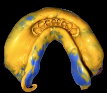
Figure 1 – Final impression
Frameworks were fabricated, verified, and adjusted. Occlusal rim was fabricated [Figure 2] and maxillo-mandibular relations were recorded [Figure 3], followed by wax try-in and occlusal adjustments. The dentures were processed, inserted, and post-insertion instructions were provided [Figures 4a-4e].
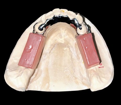
Figure 2 – Occlusal rim
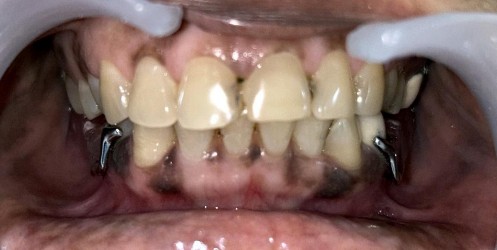
Figure 3 – Jaw relation record
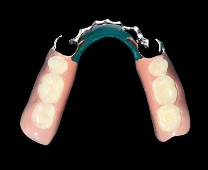
Figure 4a – Final cast partial denture (occlusal surface)
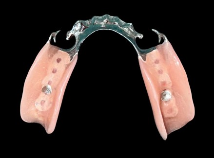
Figure 4b – Final cast partial denture (intaglio surface)
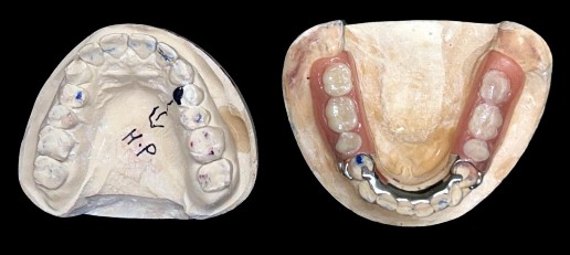
Figure 4c – Final cast partial denture on the mandibular cast
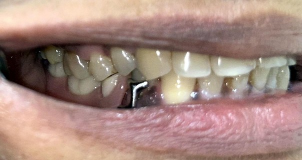
Figure 4d – Final cast partial denture in the patient’s mouth (left lateral view)
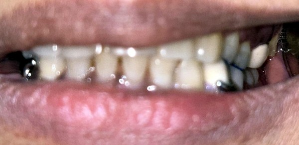
Figure 4e – Final cast partial denture in the patient’s mouth (right lateral view)
Follow-Up & Outcome
The patient was reviewed at one week, one month, and three months, reporting no discomfort and high satisfaction. At six months, she expressed excellent adaptation, improved mastication, and enhanced aesthetics.
DISCUSSION:
Cast Partial Dentures (CPDs) are the preferred choice over conventional fixed prostheses for Kennedy Class I cases due to the lack of posterior abutments.13 In the present clinical scenario, the patient opted for a cost-effective treatment, making a conventional CPD suitable for bilateral distal extension in the mandibular arch. Implant-supported fixed prostheses were not considered due to their lengthy treatment duration, high cost, inadequate bone height and width, and the patient's financial limitations.14 Prosthodontic rehabilitation for partially edentulous patients has advanced significantly with developments in CAD/CAM technology, precision- milled attachments, implant-supported prostheses, and improved impression materials.15 These innovations have enhanced the quality and acceptance of Removable Partial Dentures (RPDs). The choice of treatment primarily depends on the condition of the abutment teeth, which are frequently periodontally compromised.
The Altered Cast Impression Technique is widely used for maxillary and mandibular distal extension partially edentulous arches (Kennedy Class I and II).16 This technique improves retention and stability while reducing mucosal displacement under occlusal pressure. Originally described by Applegate, it is also known as the corrected-cast technique.17 A study by John B. Holmes on the "Influence of Impression Procedures and Occlusal Loading on Partial Denture Movement" found that partial denture movement due to occlusal loading is influenced by the impression technique and material used.18 Notably, the altered cast technique demonstrated the least movement upon occlusal loading at insertion, with minimal overall movement compared to other methods. The RPI concept and I-bar clasp system enhance retention against vertical displacement, with the I-bar's efficiency dependent on guide plane parallelism, which restricts displacement to the insertion path.19 The I-bar offers several advantages, including horizontal stability, arch stabilization, and improved retention due to its parallelism, which minimizes dislodgement.20 Additionally, it safeguards the tooth-tissue junction by preventing food impaction, provides metal coverage, and ensures proper reciprocation.
In distal extension cases, mesial rests are strategically positioned on the mesial aspect of the abutment teeth to direct occlusal forces onto the mucosa beneath the denture base extension and redistribute tipping forces mesially, ensuring better contact with the anterior teeth for added support. If a modification space exists, clasps should be placed on teeth adjacent to the edentulous area in a tripod configuration.21 Indirect retention is optimized by positioning indirect retainers as far as possible from the distal extension base, thereby improving leverage. While the incisor region provides the most effective location for indirect retainers, canines or premolars are often preferred due to the limited strength of incisors.22 In the mandible, the denture base itself may contribute to indirect retention. A double lingual bar resting on an unprepared lingual surface can enhance indirect retention through terminal rests. Furthermore, the reciprocal arm of a direct retainer positioned anterior to the fulcrum line can serve as an additional source of indirect retention.23
The longevity of an RPD is influenced by the forces exerted on abutment teeth, which vary due to differences in supporting tissue behavior.24 A CPD without precision attachments was selected for the mandibular arch due to the complexity and prolonged duration of alternative treatments, as well as financial constraints.25 Cobalt-chromium (Co-Cr) metal frameworks are preferred for CPDs due to their strength, thermal conductivity, and stability, offering an optimal
balance of deformation, stiffness, and elasticity.26 In contrast, acrylic dentures with stainless steel clasps tend to deform more rapidly. The first Co-Cr RPD framework created via Selective Laser Melting (SLM) was credited to Williams et al.27 Digital technology, including 3D E- models, has significantly enhanced digital design, cast evaluation, and reproducibility; however, conventional techniques remain relevant when properly diagnosed, planned, and executed.28
For this patient, a mandibular CPD was selected due to its superior retention, stability, and stress distribution. Complete dentures were avoided to prevent reduced proprioception and inadequate support, while implant-supported prostheses were not considered due to the patient's medical condition and reluctance to undergo surgery.29 Nonetheless, removable mandibular CPD presents challenges such as patient adaptation difficulties, plaque accumulation around abutment teeth leading to caries and periodontal disease, and aesthetic concerns due to visible metal components.30
In Kennedy Class I cases, CPDs remain the preferred choice since the absence of posterior abutments makes fixed bridgework impractical. For this patient, mandibular CPD was selected based on cost-effectiveness and treatment efficiency. Implant therapy was ruled out due to its prolonged duration, higher cost, insufficient bone support, and financial constraints. Stewart noted that bilateral clasps are appropriate for Class I partially edentulous arches, although they do not entirely control damaging forces, necessitating the strategic placement of indirect retainers bilaterally on canines. To reduce harmful horizontal forces on supporting teeth and the alveolar ridge, proper clasp fitting, denture insertion direction, and stabilizing arm positioning are crucial. Mesial rests on the posterior abutments help expand the arch of rotation, directing forces vertically and providing a buttressing effect.31 The properties of Co-Cr ensure an optimal balance between deformation, stiffness, and elasticity, contributing to long-term durability. Metal-based frameworks provide excellent strength and thermal conductivity, enhancing the overall experience for the patient. The RPI system, consisting of an I-bar retentive element, mesial rest, and distal proximal plate, minimizes torquing forces and directs occlusal loads parallel to the long axis of the abutments, thereby reducing lateral stress. Flexible RPDs, made from nylon-based thermoplastic materials, are particularly useful in mandibular bilateral distal extension cases where space for a lingual bar is limited. These flexible dentures offer aesthetic benefits through translucency and clear clasps while also providing strength, flexibility, biocompatibility, and eliminating monomer and metal allergens found in traditional denture materials.32
CONCLUSION
The design of distal extension removable dentures should be patient-specific, balancing the advantages and disadvantages of various components. When implants and fixed partial dentures are not viable, cast partial dentures provide stability, functionality, and biological restoration. A thorough clinical evaluation ensures optimal design selection, enhancing masticatory efficiency, aesthetics, and phonetics with minimal complexity. Proper maintenance ensures retention, stability, and periodontal health. In this case, the patient’s functional and aesthetic needs were met effectively and economically without invasive procedures.
REFERENCES
-
Kratochvil FJ. Influence of occlusal rest position and clasp design on movement of abutment teeth. J Prosthet Dent. 1963; 13:114–124.
-
Krol AJ. Clasp design for extension-base removable partial dentures. J Prosthet Dent. 1973;29(4):408–415.
-
Eliason CM. RPA clasp design for distal-extension removable partial dentures. J Prosthet Dent. 1983;49(1):25–27.
-
Nazarova E, Taylor TD. The RPH clasp assembly: a simple alternative to traditional designs. J Prosthodont. 2012;21(4):331–333.
-
Hakkoum MA. New clasp assembly for distal extension removable partial dentures: the reverse RPA clasp. J Prosthodont. 2016;25(5):411–413.
-
Hajjaj MS. Modified Equipoise Clasp: an esthetic option for removable dental prostheses. J Prosthodont. 2019;28(6):724–727. doi:10.1111/jopr.12916.
-
Kukunoor S, Savadi RC, Venkata Krishnam Raju K, Kumar S. A viable treatment alternative in distal extension cases: a case report. J Indian Prosthodont Soc. 2014; 14:177-180. doi:10.1007/s13191-012-0218-6.
-
Eggbeer D, Bibb R, Williams R. The computer-aided design and rapid prototyping fabrication of removable denture frameworks. J Eng Med. 2005; 219:195-202. doi:10.1243/095441105X9372.
-
Thakare KS, Bhongade ML, Charde P, Kale S, Jaiswal P, Somnath BK, Pendor S. Genetic mapping in Papillon-Lefèvre syndrome: a report of two cases. Case Rep Dent. 2013; 2013:404120. doi:10.1155/2013/404120.
-
Godbole S, Phakhan AJ, Kale S, Dahane T. Prosthodontic considerations of speech in complete denture. IOSR J Dent Med Sci. 2016; 15:41-44.
-
Mistry R, Pisulkar SK, Borle AB, Godbole S, Mandhane R. Stability in complete dentures: an overview. IOSR J Dent Med Sci. 2018; 17:36-41.
-
Pradhan B, Joshi SP, Verma A. A viable rehabilitation approach with maxillary cast partial denture and mandibular overdenture: a case report. Journal of Nepalese Prosthodontic Society. 2019;2(1):53-57.
-
D'Souza DS, Dua P. Rehabilitation strategies for partially edentulous: prosthodontic principles and current trends. Medical Journal Armed Forces India. 2011;67(3):296- 298.
-
Daniel S, Yadav R, Kumar GA. Maxillary and mandibular distal extension rehabilitation: combination of cast partial denture with RPI system and flexible partial denture: a case report. Int J Allied Med Sci Clin Res. 2020;8(1):25-31.
-
Bhathal M, Batra J, Attresh G, Sambyal S. A review on stresses induced by removable partial dentures. Int J Contemp Dent Med Rev. 2015; 2015:050115.
-
Kazem NM, Abd ST, Dawood MA, Admon M, Sabah M. The effect of removable partial denture on periodontal health status. Global J Biotechnol Biochem. 2017;6(3):512-515.
-
Kuriakose EM, Aradya A, KN RS. Cast partial denture: a preferred choice of the patient over acrylic partial and flexible denture: a case report. IJARIIT. 2019;5(3):615-617.
-
Carr AB, McGivney GP, Brown DT. Removable Partial Prosthodontics. Missouri: Elsevier Mosby; 2005.
-
Wagner B, Kern M. Clinical evaluation of removable partial dentures 10 years after insertion: success rates, hygienic problems, and technical failures. Clin Oral Investig. 2000;4(2):74-80.
-
Lee HE, Wu JH, Wang CH, et al. Biomechanical analysis of distal extension removable partial dentures with different retainers. J Dent Sci. 2008; 3:133-139.
-
Boçari G, Hysenaj N, Boçari A. The advantages of partial dentures made of cast framework cr-co toward those made of resin-based dentures. Int J Dent Sci Res. 2014;2(2):32-35.
-
Tawse-Smith A, Rivillas CC, Orozco PS, Diaz JE, Pack AR. Clinical effects of removable acrylic appliance design on gingival tissues: a short-term study. J Int Acad Periodontol. 2001;3(1):22-27.
-
Balwani TR, Dubey SG, Sathe S, Chandak A. Demystifying the role of phonetics in complete denture. Int J Res Pharm Sci. 2020; 11:2037-2043.
-
Watson CL, Reeve PE, Barnes E, Lane AE, Bates JF. The role of personality in the management of partial dentures. J Oral Rehabil. 1986; 13:83-91. doi:10.1111/j.1365-
2842. 1986.tb01558. x.
-
Runov J, Kroone H, Stoltze K, Maeda T, El Ghamrawy E, Brill N. Host response to two different designs of minor connector. J Oral Rehabil. 1980; 7:147-153. doi:10.1111/j.1365-2842. 1980.tb00431. x.
-
Al-Jammali ZM, Saad Z, Saad M. Prosthodontics clinical cases (patients with decreased inter-arch distance: case report). IJMRA. 2020; 3:1-9.
-
Crum RJ, Rooney GE Jr. Alveolar bone loss in overdentures: a 5-year study. J Prosthet Dent. 1978; 40:610-613. doi:10.1016/0022-3913(78)90054-9.
-
Rissin L, House JE, Manly RS, Kapur KK. Clinical comparison of masticatory performance and electromyographic activity of patients with complete dentures, overdentures, and natural teeth. J Prosthet Dent. 1978; 39:508-511. doi:10.1016/S0022- 3913(78)80181-4.
-
Grieder A, Cinotti WR. Periodontal Prosthesis. Louis, MO: Mosby; 1968.
-
Phoenix RD, Cagna DR, DeFreest CF. Stewart's Clinical Removable Partial Prosthodontics. Chicago: Quintessence; 2003.
-
Henderson D, McCracken WL, McGivney GP, Castleberry DJ. McCracken's Removable Partial Prosthodontics. St. Louis, MO: Mosby; 1985.
-
Singh L, Kaira SL, Dayakara HR, Singh R. Flexible denture for partially edentulous arches: a case report. J Dentofacial Sci. 2012; 1:39-42.